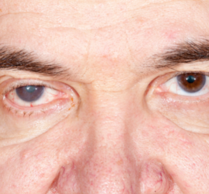


Retinal layer of the eye consists of nerve cells which transmits the light coming from outside to the brain. If the eye is like a camera, retina is the film. Macular area is just in the middle of this section. In the middle of the macula, there is the Foveo which is at the size of a pinhead where the light focuses. Foveo, also known as the yellow spot, gives the retina layer the abilities of central vision and detailed vision. Macular hole is the tear which appears right in the middle of the center of vision. A black dot appears in the vision of the patient which affects it badly. Recognizing the faces, reading newspapers or such activities are highly affected because of this condition. Since the other parts of the retina are normal, environmental vision is not affected. The most common reason of the macular hole is called “idiopatic” which is not related to anything and happens by itself. Age is somehow related to it. Other than that, traumas on the eye might also lead to macular hole.
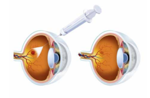
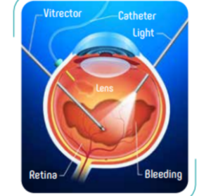
Even the factors which lead to macular hole are not exactly know, it is clear that the changes on the eye are mostly related to age. The intra ocular fluid, called Vitreous, shrinks by time, applies a pull force to the centre of sight and this force creates a hole on this area. Treatment of macular hole with medicine is not possible, the only treatment can be done is vitrectomy surgery. The vitreous fluid is cleaned, the membranes are pealed and a gas is injected into the eye. The vision might be lower after the surgery because of the gas inside the eye and the vision is corrected when the intraocular fluid replaces the gas by itself. The success rate of the operation, which means closing the macular hole is %80-90. However the rate is lower after the trauma or long-term holes. Macular hole surgery can be performed under local or general anesthesia. It is recommended to sleep on tummy for a while after the surgery to be sure that the hole is completely closed.